| Event type: | National and Regional Zoom Events |
| Date: | Tuesday 29th September 2026 |
| Time: | 11:00 am - 12:30 pm |
| Venue: | Zoom |
| Organiser: |
The Third Age Trust
|
| Cost: | Free |
| Booking: | Note that booking is required. |
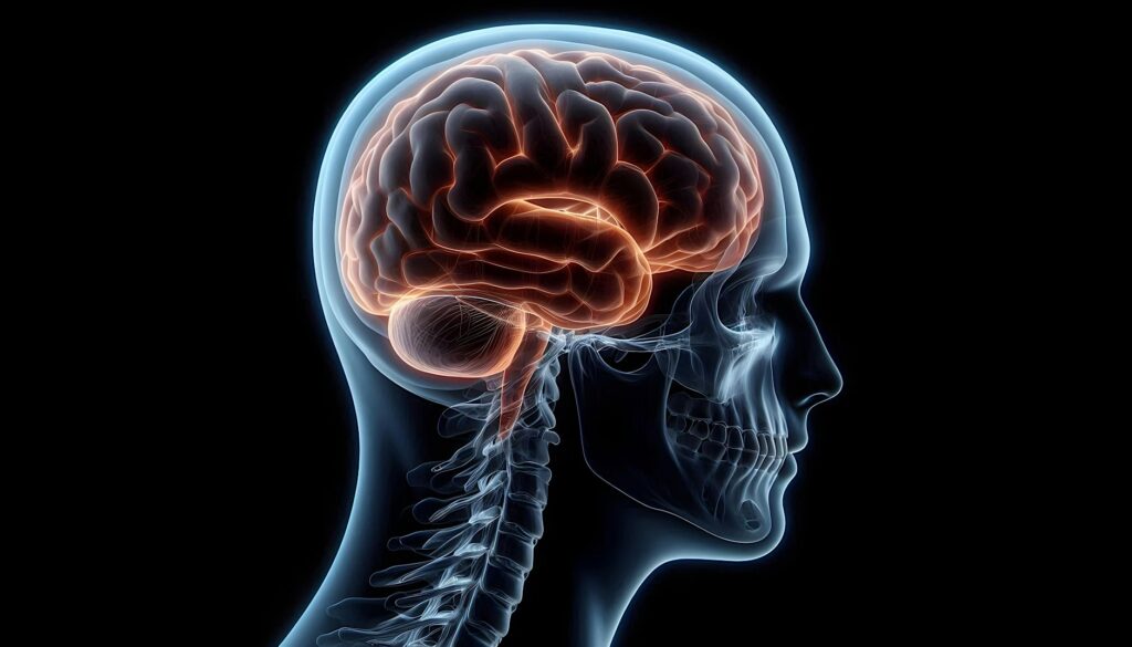
Professor Itamar Ronen describes theories of how the brain works and modern experimental approaches focussing on neuroimaging.
This event is hosted by the u3a Science Network with speaker Professor Itamar Ronen.
Since antiquity, when hypotheses on how the human body works were mostly based on theoretical constructs and religious beliefs, mankind was fascinated by the possible roles of the mysterious blob that inhabits our heads - the brain. Theories related to the nature of the brain and its functions abounded, but slowly a more grounded approach emerged - that of observational, fact-based experimental research. In its origins, experimental brain research was based on facts collected after death (post-mortem research). This provided priceless observations on brain structure, but very few insights into brain function. Insights into how the brain works started emerging with observation about loss of function - for example speech and body movement - associated with accidental brain damage. Exploration of brain function without causing brain damage was later obtained during brain surgeries, and incredibly advanced and still relevant insights into how different functions are distributed across the brain were made, but the undeniable fact remained: in order to explore brain function and physiology, one had to have physical access to the brain.
This all changed with the birth of modern medical imaging, which provided the possibility of looking at the brain (or any other organ!) without the need for physical access to it. About 130 years passed from the day when Wilhelm Roentgen published his famous X-ray images of the hand of his wife Berta, and modern biomedical imaging offers us incredible possibilities to investigate an ever-growing array of aspects of brain function and structure: from detailed anatomy to exquisite images of blood vessels, from accurate visualisation of brain activity to its chemical composition.
In the hour or so allotted to this talk, I will quickly survey how experimental brain research developed from antiquity to our days. This will set the grounds for establishing the main questions that are of interest to modern basic and clinical brain research. The second part of the talk will focus on how these questions are addressed by the main neuroimaging tools available to us today in research and in hospitals: Magnetic Resonance Imaging (MRI), Computerised X-Ray Tomography (CT) and Positron Emission Tomography (PET). Rather than explaining how these methods work, we will focus on the type of information they can provide, and on how complementary they are to each other.
About the speaker
My relationship with neuroimaging started when I was a PhD student in Tel Aviv University, where I studied chemistry with focus on the use of a method called Nuclear Magnetic Resonance (NMR, a close relative of Magnetic Resonance Imaging, or MRI) to investigate biological systems. My PhD thesis focussed on developing a NMR method that could be used to measure brain utilisation of oxygen. This project led to a collaboration with the MRI group at the University of Minnesota, one of the leading groups in MRI research and method development.
I spent the five years that followed my PhD in the University of Minnesota as a post-doctoral fellow, shifting towards developing MRI methods that would allow accurate characterisation of the microscopic structure of brain tissue, a key aspect of the brain that changes, sometimes dramatically, in disease. The multidisciplinary environment at the lab allowed me to create strong ties with neuroscientists, who played a significant role in the fact that most of my methodological research focusses on imaging the brain.
I obtained my first academic position as an Assistant Professor in the Department of Anatomy and Neurobiology at the Boston University School of Medicine. There, I participated in founding the Centre for Biomedical Imaging and together with partners from the Department of Radiology, I established a Master's degree program in Biomedical Imaging that prepared students for imaging-related work in the academia and the industry.
In 2009 I moved to the Netherlands, where I joined the MRI research centre at the Leiden University Medical Centre as Associate Professor in the Department of Radiology. I dedicated most of my research endeavours in developing methods that probe the brain chemical composition (neurochemistry) and also allow probing cellular structural features, based on the mobility of certain molecules that reside in specific brain cell types. This led me to collaborate with a large number of clinical researchers across the globe who were interested in applying this method to research the diseases in which they were interested, including Multiple Sclerosis, Lupus, Alzheimer's disease, Amyotrophic Lateral Sclerosis (ALS) and more. Included was also the collaboration with the group at the Brighton and Sussex Medical School, who were interested in an experimental model of brain inflammation (neuroinflammation). That was my first visit to Brighton, and one of the reasons I chose to move to the UK when the opportunity arose.
In 2021 I moved to Brighton, where I am now the Academic Co-Director of the Clinical Imaging Science Centre (CISC) and Chair in Medical Physics at the Brighton and Sussex Medical School (BSMS). Here I collaborate with researchers from BSMS, University of Sussex and other researchers nationwide on the topic I described earlier, as well as developing a new line of research: development and application of imaging methods for low-cost, portable MRI systems that will help (hopefully!) democratise imaging across communities and countries.